


-
1
-
2
-
3
-
4
-
5
-
6
-
7
-
8
-
9
-
10
-
11
-
12
-
13
-
14
-
15
-
16
-
17
-
18
-
19
해당 자료는 6페이지 까지만 미리보기를 제공합니다.
6페이지 이후부터 다운로드 후 확인할 수 있습니다.
6페이지 이후부터 다운로드 후 확인할 수 있습니다.


목차
Clinical and histopathological manifestations
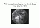
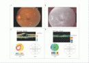
A fluorescein angiogram of the left eye in a pt with PDR
Current appoaches to prevetion and treatment
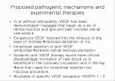
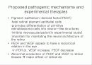
Proposed pathogenic mechanisms and experimental therapies
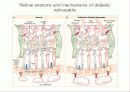
Retinal anatomy and mechanisms of diabetic retinopathy
Proposed pathogenic mechanisms and experimental therapies
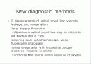
New diagnostic methods
Conclusion
A fluorescein angiogram of the left eye in a pt with PDR
Current appoaches to prevetion and treatment
Proposed pathogenic mechanisms and experimental therapies
Retinal anatomy and mechanisms of diabetic retinopathy
Proposed pathogenic mechanisms and experimental therapies
New diagnostic methods
Conclusion
본문내용
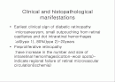
Clinical and histopathological manifestations
Earliest clinical sign of diabetic retinopathy
:microaneurysm, small outpouching from retinal capillaries and dot intraretinal hemorrhages
:all(type 1), 80%(type 2)-20years
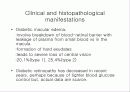
Preproliferative retinopathy
:have increase in the number and size of intraretinal hemorrhages(cotton-wool spots)- indicate regional failure of retinal microvascular circulation(ischemia)
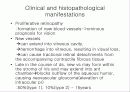
Clinical and histopathological manifestations
Proliferative retinopathy
:formation of new blood vessels->ominous prognosis for vision
New vessels
can extend into vitreous cavity,
hemorrhage into vitreous, resulting in visual loss,
can cause tractional retinal detachments from the accompanying contractile fibrous tissue
Late in the course of ds, new vs may form within the stroma of iris and may extend into ant chamberblocks outflow of the aqueous humor, causing neovascular gloucoma(elevation of intraocular pr)
:50%(type 1), 10%(type 2) - 15years
Earliest clinical sign of diabetic retinopathy
:microaneurysm, small outpouching from retinal capillaries and dot intraretinal hemorrhages
:all(type 1), 80%(type 2)-20years
Preproliferative retinopathy
:have increase in the number and size of intraretinal hemorrhages(cotton-wool spots)- indicate regional failure of retinal microvascular circulation(ischemia)
Clinical and histopathological manifestations
Proliferative retinopathy
:formation of new blood vessels->ominous prognosis for vision
New vessels
can extend into vitreous cavity,
hemorrhage into vitreous, resulting in visual loss,
can cause tractional retinal detachments from the accompanying contractile fibrous tissue
Late in the course of ds, new vs may form within the stroma of iris and may extend into ant chamberblocks outflow of the aqueous humor, causing neovascular gloucoma(elevation of intraocular pr)
:50%(type 1), 10%(type 2) - 15years
추천자료
- 가격5,000원
- 페이지수19페이지
- 등록일2006.10.20
- 저작시기2005.1
- 파일형식파워포인트(ppt)
- 자료번호#367834

본 자료는 최근 2주간 다운받은 회원이 없습니다.


소개글