


-
1
-
2
-
3
-
4
-
5
-
6
-
7
-
8
-
9
-
10
-
11
-
12
-
13
-
14
-
15
-
16
-
17
-
18
-
19
-
20
-
21
-
22
해당 자료는 7페이지 까지만 미리보기를 제공합니다.
7페이지 이후부터 다운로드 후 확인할 수 있습니다.
7페이지 이후부터 다운로드 후 확인할 수 있습니다.


목차
뇌종양
신경교종
-신경교종의 분류
교모세포종
정의
발생부위
원인
증상
진단
치료
재발 및 전이
간호
수술 전후 간호
case study
신경교종
-신경교종의 분류
교모세포종
정의
발생부위
원인
증상
진단
치료
재발 및 전이
간호
수술 전후 간호
case study
본문내용
idone and draped in usual fashion.
Under the navigation system, tumor margin was confirmed and incision line and craniotomy margin was decided. A bicoronal scalp incision was made 2 cm behine hair line. After reflection of scalp flap downward, three key holes were made and they were connected with Midas air dirll. Bone flap was removed and dural bleeding was controlled with bipolar coagulator. After dura tenting suture was done. Tumor margin was rechecked with navigation system. Dura was incised cruciately. After appling the multiple dura stay suture. Cortex was coagulated with bipolar coagulator.
Under the surgical microscope, internal decompression ans extrapedal removal of tumor was performed. Tumor was poorly demarkated. The tumor had much blood. And it was removed gross totally. Some bleeding was controlled with bipolar coagulator. The operative field was covered with surgicel. There was no oozing and active bleeding. The dura was closed watertightly. The bone flap was fixed to previous site with screws and plates.
After meticulous bleeding control, the wound was closed in layer by layer.
The patient tolerated above procedure well.
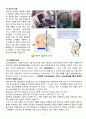
※ Brain CT CE (2012.11.21.) (Post OP)
S/P resection of the preexisting brain tumor in the right frontal lobe.
Smal amount of marginal hemorrhage.
Postop. pneumocephalus.
★ Brain, right frontal, biopsy;
Glioblastoma (WHO grade IV)
The result of immunohistochemistry;
1) GFAP: focal positive for tumor cells
2) NF: negative for tumor cells
3) p53: positive up to 95% of tumor cells
4) Ki-67: positive up to 70% of tumor cells
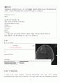
※ Brain MRI (2012.11.23.)
1. S/P resection of brain tumor in the right frontal lobe.
2. Focal residual enhancing lesion at the posteroinferior portion of
the surgical margin (about 8mm in size).
--> Suspicious focal residual tumor
--> REC.) regular follow-up MRI
3. Small amount of marginal hemorrhage with subtle thin enhancement
along the surgical margin due to postop. BBB breakdown.
4. No gross interval change of the preexisting peritumoral edema in the right frontal lobe.
※ Brain CT CE(-) (2012.11.29.) (POD 8일째)
●방사선종양학과 Prof.
안녕하십니까 상기환자 B-tumor in Rt. F.로 adm.중이신분으로 Bx.상 Glioblastoma (WHO grade IV)
check되어 귀과적 management위해 의뢰드리오니 고진선처 부탁드립니다. 감사합니다.
Dx: Rt brain tumor(frontal), postop
Pathology & Stage: glioblastoma(grade IV)
OP or Chemo: craniotomy & tumorectomy(2012. 11/21)
- 상기 환자 면담 및 진찰 했으며 다음과 같이 방사선치료 진행하겠습니다 -
면담 및 진찰 : 2012. 11/28
CT Scan & simulation : 12/03
치료시작 : 12/10
치료부위(RT Field) : brain
총선량 및 치료횟수(TDF) : 2Gy x 32fxs = >64Gy/7wks 감사합니다.
※ 항암제 용량의 결정
1) 기준: 체표면적 (body surface area, BSA)
2) 용량 단위: mg/㎡를 사용
3) Temodal 체표면적당 75mg/㎡
Temodal = 75mg/m2
체표면적(BSA) = (신장 * 체중 / 3600) ^ 1/2
신장 163.7 몸무게 70kg
체표면적(BSA): (163.7 * 70 / 3600) ^ 1/2 = 1.59
투영용량: 1.59 * 75 = 119
참고자료
이향련외(2007), 성인간호학 Ⅱ, 수문사 : 서울
서문자외(2004), 성인간호학 Ⅱ 현문사 : 서울
신경외과학(2005), 대한신경외과학회
김명자외(2005), 기본간호학(상)(하), 현문사 : 서울
5. 서울대학교병원 사이트
6. 국림암센터 사이트
7. 국가암정보센터 사이트
8. 드러그인포 사이트
8. http://cafe.naver.com/glioblas
Under the navigation system, tumor margin was confirmed and incision line and craniotomy margin was decided. A bicoronal scalp incision was made 2 cm behine hair line. After reflection of scalp flap downward, three key holes were made and they were connected with Midas air dirll. Bone flap was removed and dural bleeding was controlled with bipolar coagulator. After dura tenting suture was done. Tumor margin was rechecked with navigation system. Dura was incised cruciately. After appling the multiple dura stay suture. Cortex was coagulated with bipolar coagulator.
Under the surgical microscope, internal decompression ans extrapedal removal of tumor was performed. Tumor was poorly demarkated. The tumor had much blood. And it was removed gross totally. Some bleeding was controlled with bipolar coagulator. The operative field was covered with surgicel. There was no oozing and active bleeding. The dura was closed watertightly. The bone flap was fixed to previous site with screws and plates.
After meticulous bleeding control, the wound was closed in layer by layer.
The patient tolerated above procedure well.
※ Brain CT CE (2012.11.21.) (Post OP)
S/P resection of the preexisting brain tumor in the right frontal lobe.
Smal amount of marginal hemorrhage.
Postop. pneumocephalus.
★ Brain, right frontal, biopsy;
Glioblastoma (WHO grade IV)
The result of immunohistochemistry;
1) GFAP: focal positive for tumor cells
2) NF: negative for tumor cells
3) p53: positive up to 95% of tumor cells
4) Ki-67: positive up to 70% of tumor cells
※ Brain MRI (2012.11.23.)
1. S/P resection of brain tumor in the right frontal lobe.
2. Focal residual enhancing lesion at the posteroinferior portion of
the surgical margin (about 8mm in size).
--> Suspicious focal residual tumor
--> REC.) regular follow-up MRI
3. Small amount of marginal hemorrhage with subtle thin enhancement
along the surgical margin due to postop. BBB breakdown.
4. No gross interval change of the preexisting peritumoral edema in the right frontal lobe.
※ Brain CT CE(-) (2012.11.29.) (POD 8일째)
●방사선종양학과 Prof.
안녕하십니까 상기환자 B-tumor in Rt. F.로 adm.중이신분으로 Bx.상 Glioblastoma (WHO grade IV)
check되어 귀과적 management위해 의뢰드리오니 고진선처 부탁드립니다. 감사합니다.
Dx: Rt brain tumor(frontal), postop
Pathology & Stage: glioblastoma(grade IV)
OP or Chemo: craniotomy & tumorectomy(2012. 11/21)
- 상기 환자 면담 및 진찰 했으며 다음과 같이 방사선치료 진행하겠습니다 -
면담 및 진찰 : 2012. 11/28
CT Scan & simulation : 12/03
치료시작 : 12/10
치료부위(RT Field) : brain
총선량 및 치료횟수(TDF) : 2Gy x 32fxs = >64Gy/7wks 감사합니다.
※ 항암제 용량의 결정
1) 기준: 체표면적 (body surface area, BSA)
2) 용량 단위: mg/㎡를 사용
3) Temodal 체표면적당 75mg/㎡
Temodal = 75mg/m2
체표면적(BSA) = (신장 * 체중 / 3600) ^ 1/2
신장 163.7 몸무게 70kg
체표면적(BSA): (163.7 * 70 / 3600) ^ 1/2 = 1.59
투영용량: 1.59 * 75 = 119
참고자료
이향련외(2007), 성인간호학 Ⅱ, 수문사 : 서울
서문자외(2004), 성인간호학 Ⅱ 현문사 : 서울
신경외과학(2005), 대한신경외과학회
김명자외(2005), 기본간호학(상)(하), 현문사 : 서울
5. 서울대학교병원 사이트
6. 국림암센터 사이트
7. 국가암정보센터 사이트
8. 드러그인포 사이트
8. http://cafe.naver.com/glioblas
키워드
추천자료
- 가격3,300원
- 페이지수22페이지
- 등록일2014.05.16
- 저작시기2014.3
- 파일형식한글(hwp)
- 자료번호#917870

본 자료는 최근 2주간 다운받은 회원이 없습니다.


소개글