


-
1
-
2
-
3
-
4
-
5
-
6
-
7
해당 자료는 2페이지 까지만 미리보기를 제공합니다.
2페이지 이후부터 다운로드 후 확인할 수 있습니다.
2페이지 이후부터 다운로드 후 확인할 수 있습니다.


본문내용
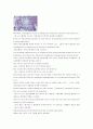
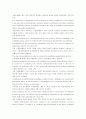
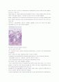
그림 4-5 심상성 천포창
An intraepidermal suprabasal cleft is visible that has resulted from suprabasal acantholysis.
표피안 기저위의 틈은 보여서 기저위 극세포융해증으로부터 유래된다.
It contains acantholytic and inflammatory cells.
그것은 극세포융해와 염증세포를 포함한다.
Acantholysis occurs in a number of different pathologic processes that do not have a uniform etiology and pathogenesis.
극세포융해는 많은 다른 병리적인 경과에서 나타나서 단일한 병인과 병인형성을 가지지 않는다.
It is important to distinguish between diseases in which acantholysis is the primary event and leads to intraepidermal cavitation (primary acantholysis) and those conditions where epidermal cells are secondarily shed from the walls of established intraepidermal blisters (secondary acantholysis).
질병사이에 구분이 중요하며, 극세포융해증이 주된 사건이며 표피안의 공동화(주된 극세포융해)를 가져오고, 이런 조건들이 있는 표피 세포는 다음으로 성립된 표피내 수포의 벽으로부터 떨어지게 된다.
Primary acantholysis is a pathogenetically relevant event in diseases belonging to the pemphigus group, where it results from the interaction of autoantibodies and antigenic
determinants on the keratinocyte membranes (see Fig. 4-5) and is mediated by epidermal proteases (see Chap. 59).
주된 극세포융해증은 천포창 그룹에 속하는 질병의 병리적 유발되어 상대적인 사건이며, 거기서는 각질세포막위에서 자가항체와 자가항원 결정원의 상호작용으로부터 나오며, 표피 프로테이즈에 의하여 매개된다.
This type of acantholysis can also be produced by pemphigus autoantibodies in vitro.
각질세포융해 형태는 또한 실험관에서 천포창 자가항체에 의하여 생산된다.
Primary acantholysis also occurs in the staphylococcal scalded-skin syndrome, where it is caused by a staphylococcal exotoxin (epidermolysin) that leads to the loss of cohesion of epidermal cells of the subcorneal epidermal layers without impairing cellular integrity
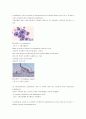
키워드
- 가격1,000원
- 페이지수7페이지
- 등록일2007.02.26
- 저작시기2007.2
- 파일형식워드(doc)
- 자료번호#396696

본 자료는 최근 2주간 다운받은 회원이 없습니다.


소개글