


-
1
-
2
-
3
-
4
-
5
-
6
-
7
-
8
-
9
-
10
-
11
-
12
-
13
-
14
-
15
-
16
-
17
-
18
-
19
-
20
-
21
-
22
-
23
-
24
-
25
-
26
-
27
-
28
-
29
-
30
-
31
-
32
해당 자료는 10페이지 까지만 미리보기를 제공합니다.
10페이지 이후부터 다운로드 후 확인할 수 있습니다.
10페이지 이후부터 다운로드 후 확인할 수 있습니다.


목차
없음
본문내용
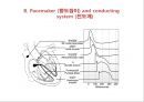
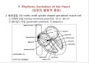
F. Rhythmic Excitation of the Heart
(심장의 율동적 흥분)
1. 전도속도 (conduction velocity)
중략
2. 동방결절 (SA node): small spindle shaped specialized muscle cell
1) 안정막 전압 (resting membrane potential): -55 to -60 mV
2) 향도잡이 전압 (pacemaker potential): 72 beats/min
3. Internodal pathways (결절간 경로)
anterior (Bachmann) nodal tract
middle (Wenckebach) nodal tract
posterior (Thorel) nodal tract
● Direction of sum vector: anterior, downward, and left
4. 방실결절 (A-V delay): more than 0.1 second
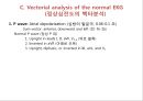
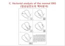
5. 좌,우 각 (Rt. & Lt. Bundle branch=Purkinje fiber)
● Direction of sum vector: downward and left
1) left to right, anterior, and toward the head
2) base to apex, posterior, and left
3) apex to base, posterior, and left 혹은 apex to base, anterior and right
● Rapid spread (0.06 sec) prevents arrhythmia - 빠른 전도가 부정맥 방지
● potential difference (전압 차이)
● extracellular recording (세포외 기록)
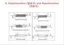
A. Depolarization (탈분극) and Repolarization (재분극)
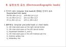
B. 심전도의 유도 (Electrocardiographic leads)
1. 표준지 유도 (Bipolar standard leads)
2. 단극지 유도 (Unipolar limb leads)와 증폭된 단극지 유도 (Augmented limb leads)
VR=RA-(RA+LA+LL) aVR=RA-(LA+LL)/2
VL=LA-(RA+LA+LL) aVL=LA-(RA+LL)/2
VF=LL-(RA+LA+LL) aVF=LL-(RA+LA)/2
3. 흉부유도 (Unipolar precordial leads or chest leads)
V1: 4th intercostal space at the right sternal border
V2: 4th intercostal space at the left sternal border
V3: Equidistant between V2 and V4
V4: 5th intercostal space in the left midclavicular line
V5: Anterior axillary line
V6: Midaxillary line
1. 전도속도 (conduction velocity)
중략
2. 동방결절 (SA node): small spindle shaped specialized muscle cell
1) 안정막 전압 (resting membrane potential): -55 to -60 mV
2) 향도잡이 전압 (pacemaker potential): 72 beats/min
3. Internodal pathways (결절간 경로)
anterior (Bachmann) nodal tract
middle (Wenckebach) nodal tract
posterior (Thorel) nodal tract
● Direction of sum vector: anterior, downward, and left
4. 방실결절 (A-V delay): more than 0.1 second
5. 좌,우 각 (Rt. & Lt. Bundle branch=Purkinje fiber)
● Direction of sum vector: downward and left
1) left to right, anterior, and toward the head
2) base to apex, posterior, and left
3) apex to base, posterior, and left 혹은 apex to base, anterior and right
● Rapid spread (0.06 sec) prevents arrhythmia - 빠른 전도가 부정맥 방지
● potential difference (전압 차이)
● extracellular recording (세포외 기록)
A. Depolarization (탈분극) and Repolarization (재분극)
B. 심전도의 유도 (Electrocardiographic leads)
1. 표준지 유도 (Bipolar standard leads)
2. 단극지 유도 (Unipolar limb leads)와 증폭된 단극지 유도 (Augmented limb leads)
VR=RA-(RA+LA+LL) aVR=RA-(LA+LL)/2
VL=LA-(RA+LA+LL) aVL=LA-(RA+LL)/2
VF=LL-(RA+LA+LL) aVF=LL-(RA+LA)/2
3. 흉부유도 (Unipolar precordial leads or chest leads)
V1: 4th intercostal space at the right sternal border
V2: 4th intercostal space at the left sternal border
V3: Equidistant between V2 and V4
V4: 5th intercostal space in the left midclavicular line
V5: Anterior axillary line
V6: Midaxillary line
키워드
추천자료
- 가격3,000원
- 페이지수32페이지
- 등록일2013.10.31
- 저작시기2010.1
- 파일형식기타(pptx)
- 자료번호#889629

본 자료는 최근 2주간 다운받은 회원이 없습니다.


소개글