


-
1
-
2
-
3
-
4
-
5
-
6
-
7
-
8
-
9
-
10
해당 자료는 3페이지 까지만 미리보기를 제공합니다.
3페이지 이후부터 다운로드 후 확인할 수 있습니다.
3페이지 이후부터 다운로드 후 확인할 수 있습니다.


목차
1.chief complaint
2.present illness
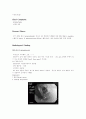
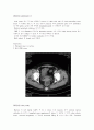
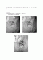
3.radiologic finding
4.lab finding
5.diagnosis
6.plan
7.disease review colorectal ca 의 종류, 병기분류,치료, 합병증, 예후
2.present illness
3.radiologic finding
4.lab finding
5.diagnosis
6.plan
7.disease review colorectal ca 의 종류, 병기분류,치료, 합병증, 예후
본문내용
nile polyp
①<10세에 호발, distal colon, rectum
②no malignant potential, cherry-red color, bleeding
3. neoplastic mucosal lesion
(1)tubular adenoma(m/c)
①>60세 호발
②15% malignant change
(특히 size >2cm, ulceration 이면)
(2)villous adenoma
①distal colon, old age
②senile type(m/c), soft consistency
③40% 악성 가능성
④Sx. : rectal bleeding, secretion
(3)tubulovillous adenoma
①villous component : 10~30%
②22% malignant potential
4. malignant tumor
(1)발생률
①증가추세
②colon (M
(2)원인
①premalignant lesion : ulcerative colitis, familial polyposis
②benzpyrene hydroxylase 는 감소 - high fat content
③high fat diet
(3)병리
①95% - adenocarcinoma, 5% - sarcoma
②호발부위 : Lt < Rt
③Genetics of colorectal carcinogenesis
(4)전파 및 전이
①direct extension
- intramural extension 특히 submucosal estension
: distal 로는 4cm 이상의 extension은 rare
잘 분화된 polypoid 면 2cm 도 good
②lymphatics - transmural 인 경우는 90% LN metastasis
wall 일부 국한시, 40% LN metastasis
- lymphatic block 생기면 poor
특히 superior hemorrhoidal artery 주위 LN
③hematogenous - rectum> lung > bone
④peritoneal seeding - ovary
⑤latogenic spread
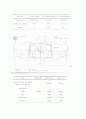
(5)TNM staging & Dukes classification
stage
Pathologic Description
Approximate 5-Year Survival, %
Duke
TNM
numeric
A
T1N0M0
Ⅰ
Cancer limited to mucosa and submucosa
>90
B1
T2N0M0
Ⅰ
Cancer extends into muscularis
87
B2
T3N0M0
Ⅱ
Cancer extends into or through serosa
70~80
C
TxN1M0
Ⅲ
Cancer involves regional lymph nodes
35~65
D
TxNxM1
Ⅳ
Distant metastases (i.e., liver, lung, etc.)
<5
TNM stage
Tumor
0 = none evident
is = In situ, limited to mucosa
1 = Invasion of submucosa
2 = Invasion of muscularis propria
3 = Invasion of subserosa or nonperitonealized pericolic fat
4 = Invasion of contiguous structures
Lymph Nodes
0 = none evident
1 = 1~3 pericolic nodes
2 = 4 or more pericolic nodes
3 = Any node along named vessel
Distant Metastasis
0 = none evident
1 = evidence of distant metastasis
(6)clinical simpton
①location
②size
③Cx. 유무에 따라 다르다.
Rt colon side
①bleeding, anemia, anrexia, indigestion, wt loss
②Rt side, mild abdominal pain, 가끔 palpable mass
triad- anemia, weakness, RLQ mass
Lt. side colon
①obstruction, bowl habit change
②loss of caliber of feces
③bloody watery stool
Rectum
bloody stool(+ mucus), tenesmus, incomplete defecation feeling
(7) 진단
①Hx & suspection, Digital rectal exam
②Sigmoidoscopy, colonofiberscope
③ba enema
④U/S, CT
(8) Bowel preparation
mechanical cleansing enema, + oral nonabsorbable antibiotics
(9)치료
수술
no touch isolation technique
①early ligation of drain vessel
②clamp of proximal & distal bowel
③minimal manipulation of cancer
합병증
①massive bleeding, obstruction, perforation - emegency op
②massive bleeding 시는 bowel cleaning effect
수술방법
①anterior resection : upper rectum lesion
②low anterior resection : mid~ lower rectum lesion (sphincter saving)
③Abdominal perineal resection (Miles' operation) : distal rectum lesion
Reference
1) Harrison's Principles of Internal Medicine 15th edition
McGraw-Hill pub 2001
2)Sabiston Textbook of Surgery 16th edition
W B Saunders pub 2001
①<10세에 호발, distal colon, rectum
②no malignant potential, cherry-red color, bleeding
3. neoplastic mucosal lesion
(1)tubular adenoma(m/c)
①>60세 호발
②15% malignant change
(특히 size >2cm, ulceration 이면)
(2)villous adenoma
①distal colon, old age
②senile type(m/c), soft consistency
③40% 악성 가능성
④Sx. : rectal bleeding, secretion
(3)tubulovillous adenoma
①villous component : 10~30%
②22% malignant potential
4. malignant tumor
(1)발생률
①증가추세
②colon (M
(2)원인
①premalignant lesion : ulcerative colitis, familial polyposis
②benzpyrene hydroxylase 는 감소 - high fat content
③high fat diet
(3)병리
①95% - adenocarcinoma, 5% - sarcoma
②호발부위 : Lt < Rt
③Genetics of colorectal carcinogenesis
(4)전파 및 전이
①direct extension
- intramural extension 특히 submucosal estension
: distal 로는 4cm 이상의 extension은 rare
잘 분화된 polypoid 면 2cm 도 good
②lymphatics - transmural 인 경우는 90% LN metastasis
wall 일부 국한시, 40% LN metastasis
- lymphatic block 생기면 poor
특히 superior hemorrhoidal artery 주위 LN
③hematogenous - rectum> lung > bone
④peritoneal seeding - ovary
⑤latogenic spread
(5)TNM staging & Dukes classification
stage
Pathologic Description
Approximate 5-Year Survival, %
Duke
TNM
numeric
A
T1N0M0
Ⅰ
Cancer limited to mucosa and submucosa
>90
B1
T2N0M0
Ⅰ
Cancer extends into muscularis
87
B2
T3N0M0
Ⅱ
Cancer extends into or through serosa
70~80
C
TxN1M0
Ⅲ
Cancer involves regional lymph nodes
35~65
D
TxNxM1
Ⅳ
Distant metastases (i.e., liver, lung, etc.)
<5
TNM stage
Tumor
0 = none evident
is = In situ, limited to mucosa
1 = Invasion of submucosa
2 = Invasion of muscularis propria
3 = Invasion of subserosa or nonperitonealized pericolic fat
4 = Invasion of contiguous structures
Lymph Nodes
0 = none evident
1 = 1~3 pericolic nodes
2 = 4 or more pericolic nodes
3 = Any node along named vessel
Distant Metastasis
0 = none evident
1 = evidence of distant metastasis
(6)clinical simpton
①location
②size
③Cx. 유무에 따라 다르다.
Rt colon side
①bleeding, anemia, anrexia, indigestion, wt loss
②Rt side, mild abdominal pain, 가끔 palpable mass
triad- anemia, weakness, RLQ mass
Lt. side colon
①obstruction, bowl habit change
②loss of caliber of feces
③bloody watery stool
Rectum
bloody stool(+ mucus), tenesmus, incomplete defecation feeling
(7) 진단
①Hx & suspection, Digital rectal exam
②Sigmoidoscopy, colonofiberscope
③ba enema
④U/S, CT
(8) Bowel preparation
mechanical cleansing enema, + oral nonabsorbable antibiotics
(9)치료
수술
no touch isolation technique
①early ligation of drain vessel
②clamp of proximal & distal bowel
③minimal manipulation of cancer
합병증
①massive bleeding, obstruction, perforation - emegency op
②massive bleeding 시는 bowel cleaning effect
수술방법
①anterior resection : upper rectum lesion
②low anterior resection : mid~ lower rectum lesion (sphincter saving)
③Abdominal perineal resection (Miles' operation) : distal rectum lesion
Reference
1) Harrison's Principles of Internal Medicine 15th edition
McGraw-Hill pub 2001
2)Sabiston Textbook of Surgery 16th edition
W B Saunders pub 2001
추천자료
- 가격1,000원
- 페이지수10페이지
- 등록일2003.11.06
- 저작시기2003.11
- 파일형식한글(hwp)
- 자료번호#231089

본 자료는 최근 2주간 다운받은 회원이 없습니다.


소개글