


-
1
-
2
-
3
-
4
-
5
-
6
-
7
-
8
-
9
-
10
-
11
해당 자료는 3페이지 까지만 미리보기를 제공합니다.
3페이지 이후부터 다운로드 후 확인할 수 있습니다.
3페이지 이후부터 다운로드 후 확인할 수 있습니다.


목차
1. 염색체 구조2. 세포분열 (cell division) 1) 유사분열(mitosis) 2) 감수분열 (meiosis, reduction division)3. 염색체 기법 (techniques of chromosome study) 1) 표본제작 방법 2) 자기방사법 (autoradiography) 3) 염색체 분염법 (differential staining methods) 4) 고정도 분염법 (high-resolution banding)4. 인체 염색체의 분류 1) 염색체의 규정 2) 인체핵형 (karyotype)
본문내용
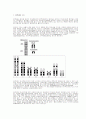
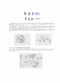
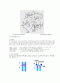
체에 따라서는 부수체가 있는 경우가 있다. 위의 기준으로 사람의 핵형은 크게 상염색체와 성염색체로 구분하여, 상염색체를 1번에서 22번까지 분류하고 이들을 다시 A군(group)에서 G군의 묶음으로 나눈다. 그리고 성염색체는 X염색체와 Y염색체로 구분한다.
① A군 : 1,2,3번의 가장 큰 3쌍의 염색체이며, 크기와 동원체의 위치에서 다른 염색체군과 쉽게 구별된다. 2차 협착이 1번 염색체의 장완 기단부(proximal portion)에 가끔 보인다.
② B군 : A군보다 짧으며, 4, 5번의 차중부(submetacentric) 염색체군이다.
③ C군 : 6번에서 12번까지의 염색체군으로서, 차중부 염색체이다. 9번 염색체는 장완의 기단부에 2차 협착이 가끔 보인다.
④ D군 : 13∼15번인 중간 크기의 차단부(acrocetric) 염색체로서, 13번 염색체는 단완에 현저한 부수체를 갖고 있으며, 14번 염색체도 단완에 작은 부수체가 있다. 이들 부수체는 항상 보이는 것은 아니다.
⑤ E군 : 16∼18번의 중부(metacentric) 또는 차중부 염색체인데, 16번 염색체는 거의 중부 염색체이며, 가끔 장완에 2차 협착이 보인다.
⑥ F군 : 19번과 20번 염색체로 거의 중부염색체이다.
⑦ G군 : 21번과 22번 염색체군인데, D군에 비해서 크기가 작은 차단부 염색체이다. 이들은 단완에 부수체를 갖고 있다.
⑧ 성염색체(sex chromosome) : X염색체와 Y염색체이다. X염색체는 상염색체군의 C군에서 7번과 8번 염색체 사이의 크기이고, 중부 염색체에 가깝다. 정상 여성은 이 X염색체를 2개 갖고 있으나 남성은 X염색체와 Y염색체를 갖고 있는데, Y염색체는 이질염색질로 형광법으로 쉽게 감별되며, G군 크기의 차단부 염색체이고 장완에 2차 협착이 나타난다. 부수체를 갖고 있지 않으며, 장완이 서로 가깝게 접해있다. 정상 남성은 X염색체와 Y염색체를 갖는 이형배우자성이다.
References
Arrighi FE, Hsu TC. Localization of heterochromatin in human chromosomes. Cytogenetics 10:81-86, 1971.
Bobrow M, Madan K, Pearson, PL. Staining of some specific regions of human chromosomes, particularly the secondary constriction of No. 9. Nature New Biol. 238:122-124, 1972.
Bobrow M, Madan K. The effects of various banding procedures on human chromosomes studied with acridine orange. Cytogenetics and Cell Genetics12:145-156, 1973.
Caspersson T, Zech L, Johansson C. Differential banding of alkylating fluorochromes in human chromosomes. Exp Cell Res. 60:315-319, 1970.
Dev VG; Warburton D, Miller OJ, Miller DA, Erlanger BF, Beiser SM. Consistent pattern of binding of anti-adenosine antibodies to human metaphase chromosomes. Exp Cell Res. 74:288-293, 1972.
Dutrillaux B, Lejeune J. Sur une novelle technique d'analyse du caryotype human. C R Acad Sci Paris 272:2638-2640, 1971.
Dutrillaux B. Nouveau systeme de marquage chromosomique: Les bands T. Chromosoma 41:395-402, 1973.
Howell WM, Denton TE, Diamond JR. Differential staining of the satellite regions of human acrocentric chromosomes. Experientia 31:260-262, 1975.
Latt SA. Microfluorometric detection of deoxyribonucleic acid replication in human metaphase chromosomes. Proc Nat Acad Sci. US 70:3395-3399, 1973.
Sahastrabuddhe CG, Pathak S, Hsu TC. Responses of mammalian metaphase chromosomes to endonuclease digestion. Chromosoma 69:331-338, 1978.
Schweizer D, Ambros P, Andrle M. Modification of DAPI banding on human chromosomes by prestaining with a DNA-binding oligopeptide, antibiotic distamycin A. Exp Cell Res. 111:327-332, 1978.
Seabright M. A reapid banding technique for human chromosomes. Lancet 2:971-972, 1971.
Shafer DA. Banding human chromosomes in culture with actinomycin D. Lancet 1:828, 1973.
Sumner AT, Evans HJ, Buckland RA. A new technique for distinguishing between human chromosomes. Nature New Biol. 232:31-32, 1971.
Yunis JJ, Sanchez O. The G-banded prophase chromosomes of man. Humangenetik 27:167-172, 1975.
추천자료
- 가격3,000원
- 페이지수11페이지
- 등록일2012.03.13
- 저작시기2008.11
- 파일형식한글(hwp)
- 자료번호#782540

본 자료는 최근 2주간 다운받은 회원이 없습니다.


소개글