


-
1
-
2
-
3
-
4
-
5
-
6
해당 자료는 2페이지 까지만 미리보기를 제공합니다.
2페이지 이후부터 다운로드 후 확인할 수 있습니다.
2페이지 이후부터 다운로드 후 확인할 수 있습니다.


목차
1. 서 론
1). 심근경색의 발생원인
2). 급성심근경색증 진단의 필요성
3). 목 적
2. 본 론
1) 심근경색의 진단방법
2) 심근경색의 전파는 보통 다음과 같은 형태의 심전도 유형을 볼 수 있다.
3. 결 론
1). 심근경색의 발생원인
2). 급성심근경색증 진단의 필요성
3). 목 적
2. 본 론
1) 심근경색의 진단방법
2) 심근경색의 전파는 보통 다음과 같은 형태의 심전도 유형을 볼 수 있다.
3. 결 론
본문내용
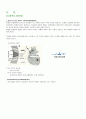
그 신티그램 (scintigram)을 취하면 접적상(hot area)으로서 경색부를 묘사할 수 있다. 동시에 심내강 신티그라피 (cardiac scintigraphy)를 행하면 경색부에 일치하여 동태가 안 좋은 곳(akinetic area)을 알아낼 수 있고 또 구출률도 측정할 수 있다. 관상동맥 종영술(coronary angiography)을 행하면 관동맥의 폐색부를 직 접 검출할 수 있으며, 좌심실 조영술로도 역시 경색부ㅗ의 동태가 안 좋은 곳을 알 수 있다.
결 론
급성심근경색증(AMI)은 발병환자의 약 30%이상이 사망하는 중증의 질병으로 과거에는 주로 50세 이상의 고연령층에서 유발되었으나 차츰 젊은층으로 확산되고 있는 추세이다. 그래서 돌연사의 위험이 따르는 만큼 심근경색의 진단까지의 시간을 줄이는 것이 주요 목적이 될 것이다.
This pictures shows how the coronary arteries originate from the ascending aorta to supply the heart with oxygenated blood. The demand for fresh, oxygenated blood is substantial for the heart. Like brain cells, the cells that work collectively to form the structure of the heart will not live for very long (respirate anaerobically) without oxygen. Coronary artery disease (CAD) results from occluded coronary arteries. The heart becomes starved for oxygen and other nutrients, and eventually stops beating. A coronary artery bypass graft operation (CABG) is performed by attaching a vein taken from the individual's leg to the ascending aorta. The other end of the vein is grafted so as to increase the flow of blood to the heart by substituting for the clogged coronary artery
키워드
추천자료
 심근경색 치료방법 중 혈전용해술에 관하여
 [관상동맥질환]협심증과 심근경색질환
- 심전도와 협심증 심근경색
- <파워포인트> 심근경색
- 심근경색환자재활둥동 및 관리
- 심근경색에 대하여
- MI(심근경색) CASE STUDY
- 케이스 스터디(Case Study) - 심근경색 (MI ; myocardial infarction)
- <심근경색이란 무엇인가>!!!
 NSTEMI 심근경색 컨퍼런스
- [재활간호][실어증환자][관절염환자][뇌졸중환자][유방암]재활간호의 배경, 재활간호의 과정,...
- 간호학과- 심근경색 문헌고찰, 의학용어
- STEMI, 심근경색증 케이스 (간호사정, 검사결과, 간호진단, 문헌고찰, 간호계획, 약물조사포함)
- 간호,case study,성인(응급실),심근경색 myocardial infarction
- 가격1,300원
- 페이지수6페이지
- 등록일2002.03.07
- 저작시기2002.03
- 파일형식한글(hwp)
- 자료번호#191703

본 자료는 최근 2주간 다운받은 회원이 없습니다.